Figure 1.

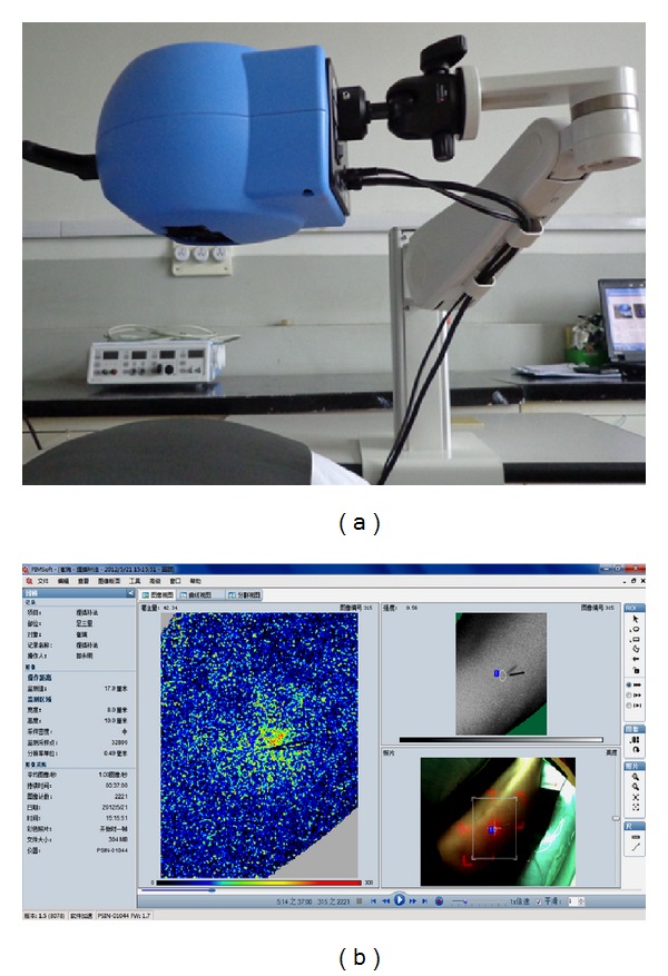
(a) Pericam Perfusion Speckle Imager (PSI). (b) The blood perfusion image of the monitor area. The brighter area is the definition of the Region of Interest (0.5 cm around the Zusanli acupoint).

(a) Pericam Perfusion Speckle Imager (PSI). (b) The blood perfusion image of the monitor area. The brighter area is the definition of the Region of Interest (0.5 cm around the Zusanli acupoint).