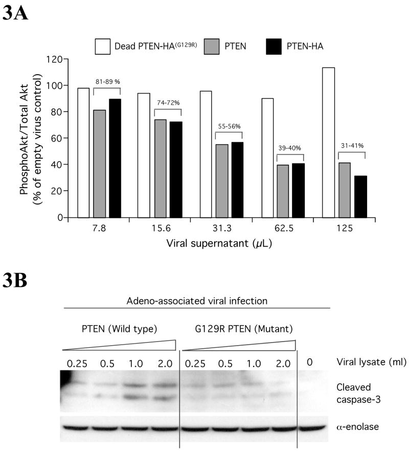
Figure 3. PTEN regulates apoptosis by inhibiting Akt activity in melanomas.
3A PTEN expression decreases pAkt levels in melanoma ccells lacking functional PTEN. UACC 903 melanoma cells lacking functional PTEN protein were infected with increasing amounts (7.8, 15.6, 31.3, 62.5, 125 μL) of adeno-associated viral constructs containing Wt PTEN, HA tagged Wt PTEN and a catalytically inactive G129R PTEN mutation. Three days later, cell lysates were collected and proteins analyzed by Western blotting. Levels of pAkt and total Akt were quantitated by densitometry and the pAkt/totalAkt ratio represented against amount of viral supernatant used for infection. Data show that pAkt expression decreases with increasing virus amount indicating PTEN reduces pAkt in melanomas (Stahl et al., 2003). 3B. PTEN triggers apoptosis in melanomas. Levels of cleaved caspase-3, an indicator of apoptosis, were measured using Western blotting following expression of viral introduced PTEN into UACC 903 melanoma cells lacking functional PTEN protein. Compared to functionally inactive G129R PTEN, wild type PTEN expression increased levels of cleaved caspase-3 indicating elevatedlevels of apoptosis. α-enolase served as control for protein loading (Stahl et al., 2003).

