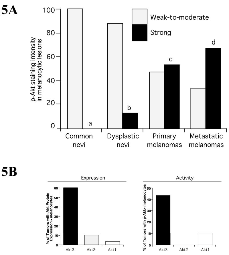
Figure 5. Akt3 expression and activity increase during melanoma development.
5A Expression of pAkt in nevi, dysplastic nevi, primary and metastatic melanoma lesions was quantitated from histological sections and intensity scored using immunohistochemical staining. Compared to common nevi that had only weak-to-moderate pAkt staining, increasing percentages of dysplastic nevi, primary and metastatic melanomas contained higher levels of Akt activity with 67% of advanced tumors having elevated activity (Stahl et al., 2004). 5B. Expression and activity of Akt isoforms (Akt1, Akt2 and Akt3) was measured in flash frozen melanoma patient tumor samples by Western blotting. Approximattely 60% of melanoma tumors expressed elevated levels of Akt3 protein compared to normal human melanocytes. Furthermore, ~43% of tumor had elevated Akt3 activity.

