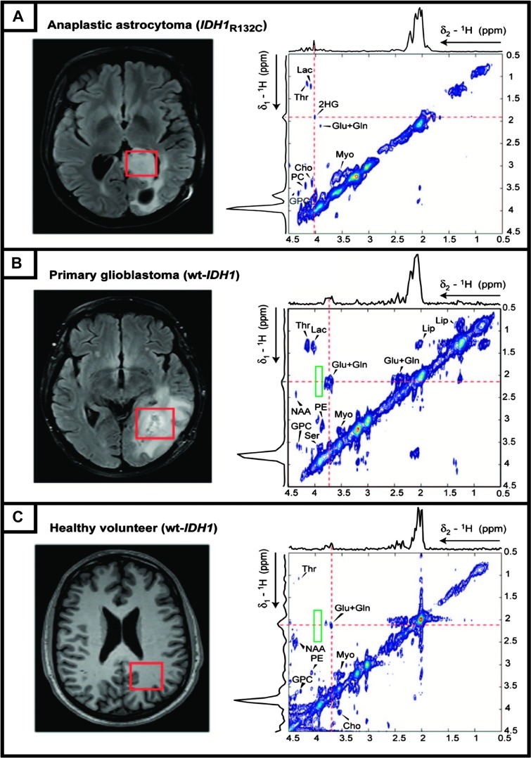
Figure 3.
In vivo 2D MRS spectra of human brain subjects acquired at 3 T. Single voxels (red rectangles, 3 x 3 x 3 cm3) are placed on the basis of fluid attenuated inversion recovery images prescribing abnormalities in tumor patients (left column), and the corresponding metabolite cross-peak are depicted as contour maps (right column). (A) Astrocytoma patient with IDH1-R132C; the Hα-Hβ cross-peak of 2-HG located at 4.02/1.91 ppm (δ2/δ1). The 2D spectra acquired (voxel size of 3.5 x 3.5 x 3.5 cm3) from a primary glioblastoma patient with wt-IDH1 (B) and healthy volunteer with wt-IDH1 (C) do not contain any 2-HG cross-peak (outlined by the green rectangle). All 2D spectra were acquired using a developed 2D localized adiabatic selective refocusing-COSY sequence with a repetition time of 45 milliseconds, 64 increments in F1 direction, 8 averages per F1 transient, and a total acquisition time of 12.8 minutes. Adiabatic pulses improve the sequence performance by providing sharp and uniform excitation slices and a robust flip angle and by significantly decreasing the chemical shift displacement error (see [31]). Adapted with permission from [15].

