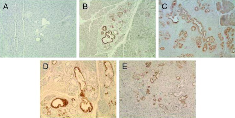
Figure 2.
Immunochemical staining of sections from wild-type (WT, A) and KPC mice with PanIN1B-2 (B) and PanIN2/3 (C) lesions and carcinoma (D, E) using anti-LCN2 antibody (AF 1857 [23]; R&D Systems, 1:1000 dilution). Strong, positive (brown) staining is apparent in the ductal lesions, whereas the normal/wild-type tissue is only weakly stained. For immunolabeling, formalin-fixed and paraffin-embedded archived tumor samples and corresponding normal tissues were stained as previously described [19]. Briefly, slides were heated to 60°C for 1 hour, deparaffinized using xylene, and hydrated by a graded series of ethanol washes. Antigen retrieval was accomplished by microwave heating in 10mM sodium citrate buffer of pH 6.0 for 10 minutes. For immunohistochemistry, endogenous peroxidase activity was quenched by 10-minute incubation in 3% H2O2. Nonspecific binding was blocked with 10%serum. Sections were then probed with lipocalin antibody (R&D Systems; AF1857) affinity-purified polyclonal goat IgG (1:1000) overnight at 4°C. Bound antibodies were detected using the avidinbiotin complex peroxidase method (ABC Elite Kit; Vector Labs, Burlingame, CA). Final staining was developed with the Sigma FAST DAB Peroxidase Substrate Kit (Sigma, Deisenhofen, Germany).

