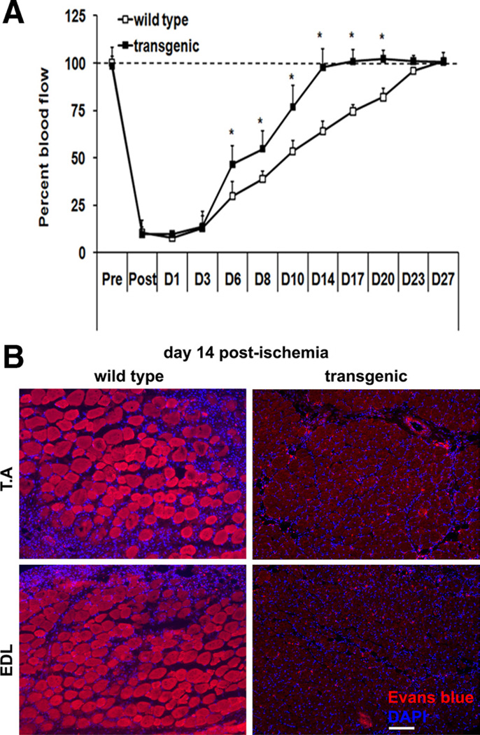
Figure 4. Time course (A) of blood perfusion.
Blood perfusion in both the ischemic and contralateral tibialis anterior (TA) was measured using laser Doppler probe. Blood perfusion in ischemic TA muscle is calculated as the percent of perfusion in the contralateral control muscle (N=14). *Statistically significant difference between wild-type and transgenic mice (P<0.001, two-way analysis of variance with repeated measurements on time). B, Representative immunofluorescent images depicting Evans blue dye staining in the ischemic TA and extensor digitorum longus (EDL) muscles from wild-type and transgenic mice on day 14 postischemia. Inclusion of Evans blue dye within a fiber is indicative of sarcolemmal damage and myofiber degeneration. Scale, 100 µm. Similar results were obtained from N=5 animals per group.

