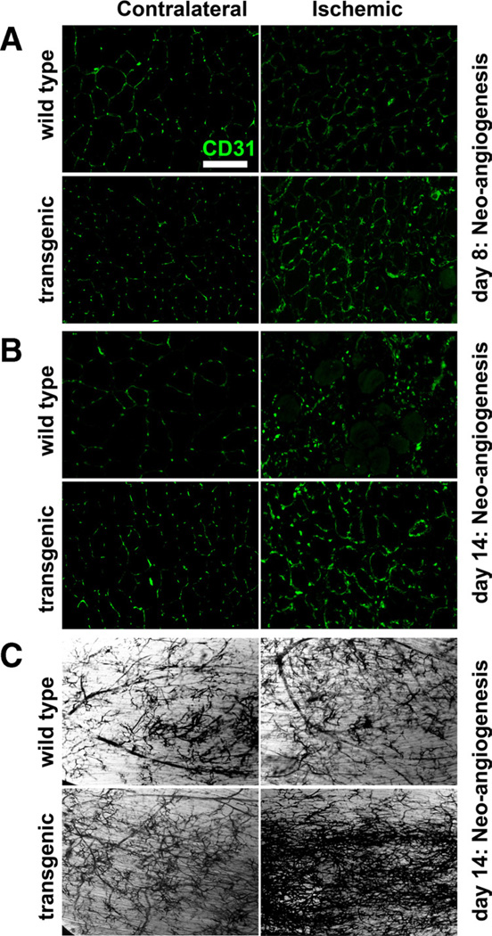
Figure 5. Neoangiogenesis in the ischemic muscle.
Representative images for the CD31 stained capillaries in contralateral and ischemic tibialis anterior (TA) cryosections from wild-type and transgenic mice on day 8 (A) and day 14 (B) postischemia. Scale, 100 µm. C, Microfil pigment vascular mapping in whole-mounted TA muscles from wild-type and transgenic mice on day 14 postischemia indicating neoangiogenesis. Similar results were obtained from N=3 to 8 animals per group.

