
Figure 6. Moderately elevated expression of PINCH rescues muscle detachment defects of the flw7 mutant.
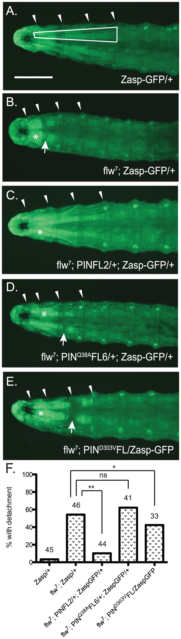
A–E) Representative images of four-day-old larvae of the indicated genotypes are shown. Ventral view allows the VIS muscles to be rapidly scored on a dissecting microscope. One of the medial pair of VIS muscles is boxed in A. White arrowheads show the relevant segment boundaries (T1,T2,T3). Areas of hypercontraction are indicated with an asterisk. Detachments are characterized by a gap in the GFP signal (arrow) Scale bar = 1 mm. F) The graph shows the percent of total animals in which the VIS muscles exhibit detachment. Number of animals examined of each genotype, pooled from triplicate experiments, is shown above the bars. ** indicates p<0.005, * indicates p<0.05, and ns indicates p>0.05.