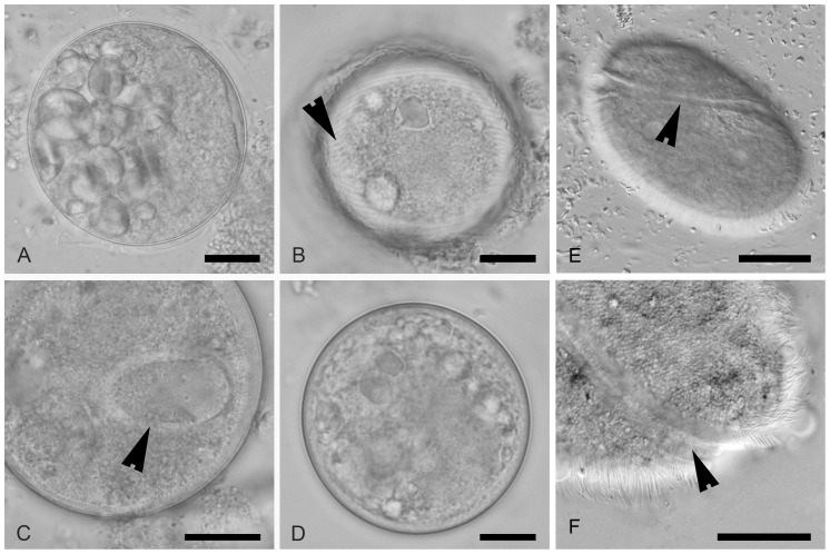
Figure 4. A–D: Comparison of cysts of Neobalantidium coli, Buxtonella sulcata and a Buxtonella-like ciliate; scale bars = 10 µm.
A. Cyst of N. coli from a domestic pig with visible ingested starch grains inside. B, D. Cysts of Buxtonella-like ciliate from an agile mangabey showing the trophozoite with visible rows of cilia (B, arrowhead). C. Cyst of B. sulcata from cattle with visible macronucleus (arrowhead). E. Trophozoite of Buxtonella sulcata with typical sulcus (arrowhead); scale bar = 20 µm. F. Detail of sulcus of Buxtonella sulcata (arrowhead); scale bar = 5 µm.

