
Figure 1. Human APOBEC3 rapidly restricts vif-deleted HIV-1JR-CSF in vivo.
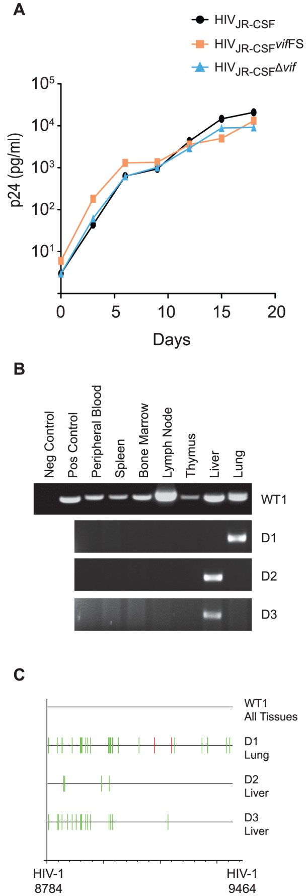
(A) Replication of HIVJR-CSF, HIVJR-CSFΔvif, and HIVJR-CSF vifFS in CEM-SS cells expressing CCR5 (CEM-SS CCR5). Culture supernatant was assayed for p24Gag by ELISA at three day intervals to determine the replication kinetics of the mutant viruses. (B) Nested PCR amplification of viral DNA from the tissues obtained one week post-exposure from a representative NSG-hu mouse infected with 9×10⁴ TCIU of wild-type HIV-1JR-CSF (WT1) or from three mice infected with 3.6×10⁵ TCIU of HIVJR-CSFΔvif (indicated as D1–3). (C) Highlighter sequence analysis of 7 wild-type and 3 Δvif HIV DNA sequences. Amplified viral DNA from panel A showed no APOBEC3 induced mutations in HIVJR-CSF (WT1 all sequence from tissues is shown together). In contrast, viral DNA from all positive tissues obtained from HIVJR-CSFΔvif infected mice had G to A (green lines) and/or C to T mutations (red lines). HIV-1JR-CSF nucleotide numbers are indicated at the bottom.