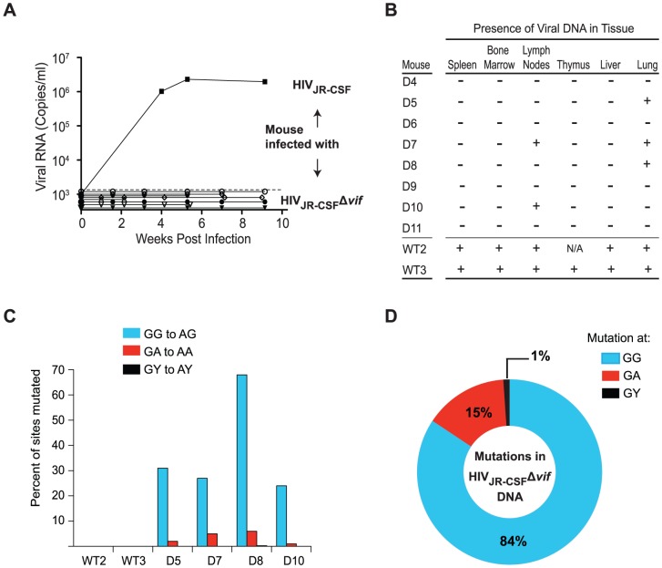
Figure 2. vif-deleted HIV-1JR-CSF does not overcome APOBEC3 restriction in vivo.
(A) Longitudinal analysis of plasma viral load in humanized mice intravenously infected with 9×10⁴ TCIU of wild-type HIV-1JR-CSF or 3.6×10⁵ TCIU of HIVJR-CSFΔvif. (B) Detection of HIV DNA (+) by nested PCR from the tissues of humanized mice in panel A. Negative tissues (−) yielded no amplified viral DNA using two independent nested PCR primer sets targeting separate regions of the viral genome. N/A = not analyzed. (C) Percentage of putative APOBEC3 mutation sites (GG, GA, GY) that were mutated in 17 viral DNA sequences amplified from the tissues of infected mice. Viral DNA from mice infected with HIVJR-CSFΔvif had 25%–65% of GG sites mutated. (D) G to A mutational profile of all viral DNA from mice infected with HIVJR-CSFΔvif. Percentages indicate the proportion of G to A mutations occurring at GG (blue), GA (red), or GY (black) sites. D4-D8, WT2-WT3, NSG-hu mice. D9-D11, NSG-BLT mice.

