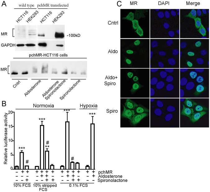
Figure 3. Human mineralocorticoid receptor can be functionally activated in HCT116 cell line.
(A, upper panel) MR expression. Whole cell lysates from wild type and pchMR-transfected HCT116 cells were analysed by western blot using anti-MR antibodies. Human kidney cells (HEK293) served as positive control. Human GAPDH was used as protein loading control. Representative fluorograms from two independent experiments giving similar results are shown (A, bottom panel) MR post-translational modifications. PchMR-transfected HCT116 cells were treated for 24 h with 3 nM aldosterone and/or 1 µM spironolactone in Mc Coy’s medium with 10% charcoal-stripped FCS. Whole cell lysates were analysed by Western blot using anti-MR antibodies. MR post-translational modifications induced by aldosterone treatment are indicated by the upward shift in the mobility of MR. A representative fluorogram from three independent experiments with superimposable results is shown (B) MR dependent luciferase activity. PcDNA3-transfected (gray bars) or pchMR-transfected (white bars) HCT116 cells were transfected with pMMTV-Luc to express firefly luciferase from an MR dependent promoter. Cell culture, aldosterone or spironolactone treatment and normoxia or hypoxia conditions are detailed in Materials and Methods section. Values of firefly luciferase activity of aldosterone-stimulated pchMR-transfected cells in 10% stripped FCS or 0.1% FCS, both in normoxic or hypoxic conditions, were compared to those of unstimulated pchMR-transfected control cells, set as 1. Values of firefly luciferase activity of pchMR-transfected cells in 10% FCS were compared to that of pcDNA3-transfected control cells, set as 1. Results were expressed as Mean± SEM (n = 4–6). **p<0.005 and ***p<0.001, vs control cells, #p<0.001 vs FCS- or aldosterone-treated cells, ANOVA followed by Bonferroni t-test or Student t-test when appropriate. (C) MR subcellular localization. PchMR-transfected HCT116 cells treated with aldosterone (3 nM) and/or spironolactone (1 µM) for 30 minutes and stained with an anti-MR antibody (green) and DAPI (blue). Images were taken with a confocal laser scanning microscope.

