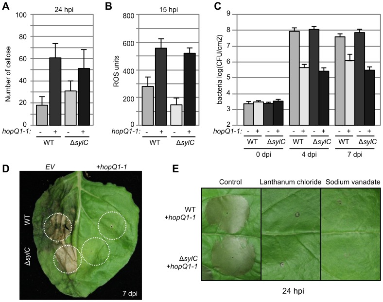
Figure 6. ΔsylC-induced immune responses are weak compared to ETI/NHR responses.
(A) Increased callose deposition by both WT and ΔsylC strains expressing the HopQ1-1 effector. Leaves were infiltrated with 2×10⁷ bacteria/mL, and fluorescent callose spots were quantified per 0.56 mm2 after aniline blue staining at 24 hpi. Error bars indicate SEM of 20 technical replicates. These experiments were repeated three times with similar results. (B) Increased oxidative burst by both WT and ΔsylC strains expressing the HopQ1-1 effector. Leaves were infiltrated with 2×10⁸ bacterial cells/mL, and reactive oxygen species (ROS) were measured at 15 hpi in leaf discs floating on 200 µM MOPS containing L-012 for 20 min. The reduced ROS level of ΔsylC(EV)-infiltrated tissue compared to WT(EV)-infiltrated tissue was due to the late time point and the fact that ROS levels are transient. Error bars indicate SEM of eight technical replicates. These experiments were repeated three times with similar results. (C) Reduced bacterial growth of both WT and ΔsylC bacteria expressing the HopQ1-1 effector. Leaves were infiltrated with 2×10⁵ bacterial cells/mL in N. benthamiana, and bacterial growth was measured at 0, 4, and 7 dpi. Error bars represent standard deviation of four biological replicates. The experiment was repeated four times with similar results. (D) HopQ1-1-expressing WT and ΔsylC bacteria do not cause symptoms when infiltrated at low densities. Leaves were infiltrated at 2×10⁵ bacterial cells/mL, and pictures were taken at 7 dpi. (E) Cell death triggered by HopQ1-1-expressing WT and ΔsylC bacteria is blocked by the calcium transport inhibitor lanthanum chloride and the ATPase inhibitor sodium vanadate. Leaves were co-infiltrated with 1×10⁸ bacteria/mL with 50 µM lanthanum chloride or 1 µM sodium vanadate, and pictures were taken at 24 hpi.

