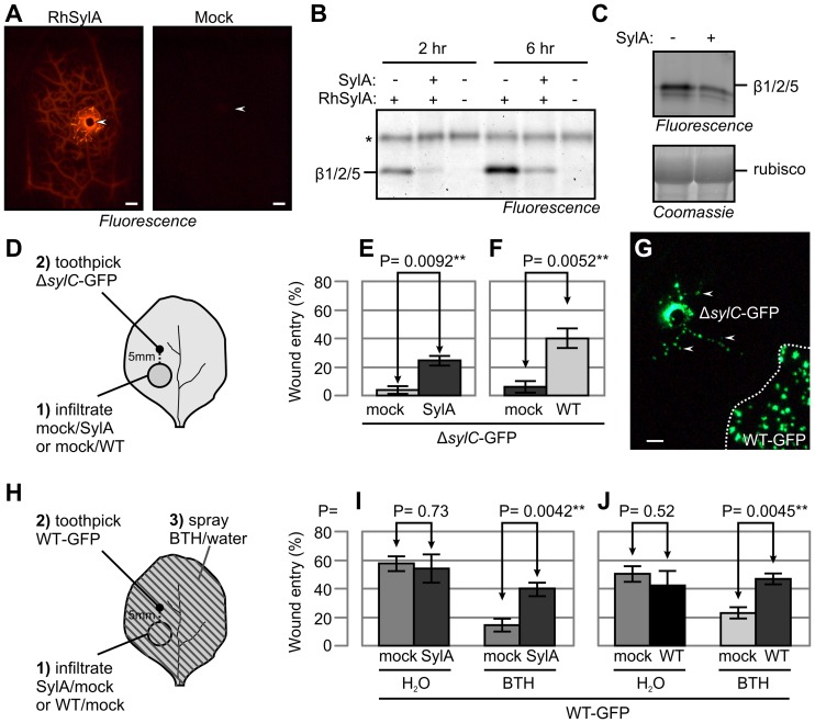
Figure 7. SylA diffuses and suppresses SA-mediated immunity in adjacent tissue.
(A) RhSylA spread through the vasculature. A 1-µl aliquot of 2 mM RhSylA or 0.1% DMSO (mock) was applied at a wound site, and a fluorescence image was taken 2 h later. Scale bar, 1 mm. Arrowheads indicate wound inoculation sites. (B) RhSylA targets the proteasome in adjacent tissue. A 1-µl aliquot of 2 mM SylA was applied to an inoculation site and preincubated for 30 min. Subsequently, 1 µl of 2 mM RhSylA was added and incubated for another 2 h or 6 h. Proteins were extracted from tissue at 1–10 mm from the application site, and labeled proteins were detected by fluorescence scanning. *, background signal. (C) SylA targets the proteasome in adjacent tissue. A 1-µl aliquot of 1 mM SylA was applied at a wound site and incubated for 4 h. The application site was removed, extracts from adjacent tissues were labeled with MVB072, and fluorescently labeled proteins were detected. (D) Procedure for assaying wound entry by ΔsylC-GFP bacteria in adjacent tissue. Leaves of WT N. benthamiana were infiltrated with 50 µM SylA and 0.25% DMSO (E), 105 WT bacteria or water (F), and the infiltrated region was marked. After 1 h (for SylA infiltration) or 1 d (for bacterial infiltration), ΔsylC-GFP bacteria were inoculated at a site 5 mm outside the infiltrated area. Wound entry was scored 5 d later by fluorescence microscopy. (E) SylA promotes wound entry by ΔsylC-GFP bacteria at a distance from the infiltrated region. (F) WT bacteria promotes wound entry at a distance from the infiltrated region. (G) Representative example of distant colonization of ΔsylC-GFP bacteria when inoculated next to areas infiltrated with WT-GFP bacteria. WT-GFP bacteria were infiltrated at 105 bacterial cells/mL (lower right, bordered by dashed line). One day later, ΔsylC-GFP bacteria were inoculated at 5 mm from the infiltrated region. The picture was taken 5 d later. WT-GFP bacteria did not spread outside the infiltrated zone, but their presence promoted wound entry by ΔsylC-GFP in adjacent tissue. Arrowheads indicate colonies of ΔsylC-GFP in tissues adjacent to the wound inoculation site. (H) Procedure for assaying adjacent colonization by WT-GFP bacteria in adjacent tissue. Leaves of WT N. benthamiana were infiltrated with 50 µM SylA, 0.25% DMSO (H), 105 WT bacteria or water (I), and the infiltrated region was marked. After 1 h (for SylA infiltration) or 1 d (for WT bacteria infiltration), WT-GFP bacteria were inoculated at 5 mm outside the infiltrated area and the plant was sprayed with 300 µM BTH or water. Wound entry was scored 5 d later by fluorescence microscopy. (I) SylA promotes wound entry in BTH-treated tissue at a distance from the infiltrated region. (J) SylA-producing WT bacteria promotes wound entry in BTH-treated tissue at a distance from the infiltrated region. (E, F, I, J) Error bars represent SEM of four independent biological replicates, each with 12 wound inoculations. P-values determined using the Student's t-test are indicated.

