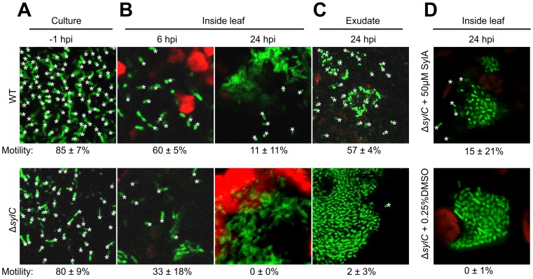
Figure 9. ΔsylC bacteria loose motility during infection.
GFP-expressing WT and ΔsylC bacteria were monitored by confocal microscopy before infiltration (A), after infiltration (B), in the exudate of infected leaves (C), and after co-infiltration with or without SylA (D). All motile bacteria are marked with a star. The percentages of motile bacteria over a 2 s timeframe are indicated on the bottom, with standard deviations for n = 5. See supplemental data for the movies and details. Bacteria are 1–2 µm long. These results are representative of three independent experiments. *, motile bacterium.

