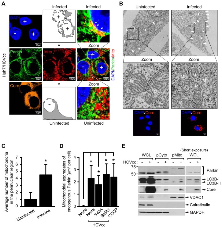
Figure 1. HCV infection induces perinuclear clustering of mitochondria and Parkin translocation to mitochondria.
(A) Representative confocal images showing perinuclear clustering of mitochondria and Parkin aggregates on the mitochondria in Huh7 cells infected with HCVcc. At 3 days post-infection, cells prestained with MitoTracker (Mito, red) were immunostained with anti-Parkin (green) and HCV core (orange) antibodies. Nuclei were stained with DAPI (blue). Infected (+) and uninfected cells (−) are marked. The illustrated images display typical distribution of mitochondria in uninfected cells and altered distribution of mitochondria in the perinuclear region of infected cells (gray). In the zoomed images, the white arrows indicate accumulation of endogenous Parkin recruited to the mitochondria in HCV-infected cells (yellow). (B) Ultrastructure of HCV-infected cells showing perinuclear clustering of damaged mitochondria. Control naïve Huh7 cells (left) and stable cells harboring HCV full-length replicon FLR-JFH1 (right) were examined by electron microscopy. In the zoomed images, typical ultrastructure of mitochondria in naïve cells and ultrastructural abnormalities of mitochondria in HCV replicon cells are shown. Organelle mark: N, nucleus; M and white arrow, mitochondria. Scale bar = 1 µM. Fluorescent images (below) indicate the expression of HCV core protein in HCV replicon cells. Cells were immunostained with anti-HCV core antibody (red). Nuclei were stained with DAPI (blue). (C) Quantification of the number of mitochondria in the perinuclear region (mean ± SEM; n≥5 cells; *p<0.05). (D) Quantification of fluorescence intensity of Parkin aggregates on the mitochondria in CCCP-treated (see Figure S1) or HCV-infected cells (A) and those treated with 3-MA or BafA1, respectively (see Figure 4A ) (mean ± SEM; n = 10 cells; *p<0.01). P values were calculated by using an unpaired Student's t-test. (E) Western blot analysis showing endogenous Parkin recruitment to mitochondria in HCV-infected cells. Huh7 cells were infected with HCVcc. At 5 days post-infection, pure cytoplasm and mitochondria fractions were isolated by ultracentrifugation as described in Materials and Methods. Cellular fractions of HCV-infected cells were analyzed by immunoblotting using antibodies specific for the indicated proteins. Fractions: whole cell lysates, WCL; purified cytoplasm, pCyto; purified mitochondria, pMito. Organelle markers: VDAC1, mitochondria; Calreticulin, ER; GAPDH, cytoplasm.

