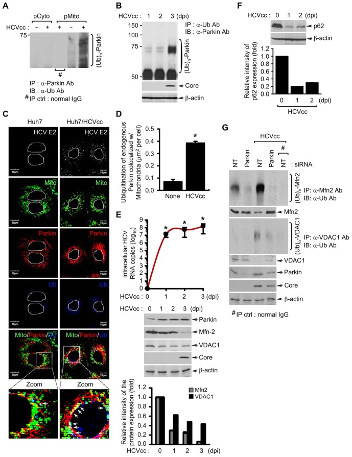
Figure 2. HCV infection induces the ubiquitination of Parkin and its mitochondrial substrates Mfn2 and VDAC1, and autophagy-associated factor p62.
(A) Parkin ubiquitination in the purified mitochondria isolated from HCV-infected cells. Parkin protein in pCyto and pMito fractions, respectively, was immunoprecipitated by anti-Parkin antibody, followed by immunoblotting with anti-ubiquitin (Ub) antibody. Normal rabbit IgG was used as a negative control for immunoprecipitation (IP). (B, E, and F) At the indicated time points post-infection, WCL were extracted from HCV-infected Huh7 cells. (B) Parkin ubiquitination in WCL extracted from HCV-infected cells. All ubiquitinated proteins were immunoprecipitated with anti-Ub antibody and analyzed by immunoblotting with anti-Parkin antibody. HCV infection was verified by immunoblotting with anti-HCV core antibody. β-actin was used as an internal loading control. (C) Confocal microscopy showing Parkin ubiquitination in HCV-infected cells. HCV-infected cells prestained with MitoTracker (Mito) were immunostained with anti-Parkin and ubiquitin (Ub) antibodies. In the zoomed images, the arrows (white spots) indicate the merge of Mito (green), Parkin (red), and Ub (blue). Nuclei are demarcated with white dot circles. Fluorescence image of HCV E2 verifies HCV-infected cells (light gray). (D) Quantitative analysis of the ubiquitination of endogenous Parkin colocalized with mitochondria in the panels (C) (mean ± SEM; n≥10 cells; *p<0.05). (E) Western blot analyses of Mfn2 and VDAC1 expression, mitochondrial substrate of Parkin, in HCV-infected cells. Intracellular HCV RNA levels were analyzed by real-time qRT-PCR, as described in Materials and Methods (mean ± SD; n = 3; *p<0.01). WCL extracted from HCV-infected cells were analyzed by immunoblotting with anti-Parkin, Mfn2, and VDAC1 antibodies, respectively. HCV infection was verified by immunoblotting with anti-HCV core antibody. β-actin was used as an internal loading control. (F) Western blot analysis of p62 expression, the autophagy-associated factor, in HCV-infected cells. WCL extracted from HCV-infected cells were analyzed by immunoblotting with anti-p62 antibody. β-actin was used as an internal loading control. (E and F) The relative intensity of Mfn2, VDAC1, and p62 expression normalized to β-actin was analyzed by ImageJ. (G) Parkin-mediated ubiquitination of Mfn2 and VDAC1 in HCV-infected cells. The ubiquitinated Mfn2 and VDAC1 proteins were analyzed by immunoprecipitation with anti-Mfn2 and VDAC1 antibodies, respectively, followed by immunoblotting with anti-Ub antibody. The protein expression levels of Parkin were analyzed by immunoblotting with anti-Parkin antibody. HCV infection was verified by immunoblotting with anti-HCV core antibody. β-actin was used as an internal loading control. Normal mouse IgG was used as a negative control for immunoprecipitation (IP). (D and E) P values were calculated by using an unpaired Student's t-test.

