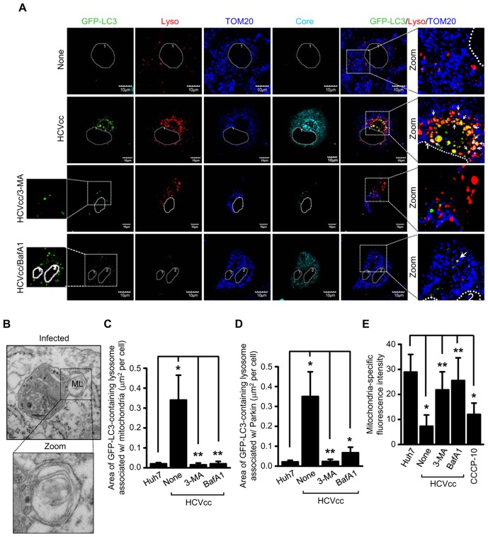
Figure 5. HCV induces complete mitophagolysosome formation.
Confocal microscopy showing the formation of mitophagolysosome in HCV-infected cells. (A) Huh7 cells transiently expressing GFP-LC3 protein were infected with HCVcc in the absence or presence of 3-MA (10 mM) and BafA1 (100 nM), respectively, for 12 h before fixation. At 3 days post-infection, cells prestained with LysoTracker (red) were immunostained with antibodies against TOM20 (blue) and HCV core (cyan). Nuclei are demarcated with white dot circles. In the zoomed images, the arrows indicate the colocalization of GFP-LC3 puncta (green), lysosome, and mitochondria in HCV-infected cells (white puncta). (B) Ultrastructure of HCV-infected cells showing the formation of mitophagolysosome. HCV FLR-JFH1 replicon cells were examined by electron microscopy. In the zoomed image, the formation of mitophagolysosome in HCV-infected cells is shown. Organelle marker: ML, mitophagolysosome. Scale bar = 0.1 µM. (C and D) Quantitative analyses of the colocalization of GFP-LC3 containing lysosomes with mitochondria (C) or Parkin (D) in the panels (A) and Figure S5 (mean ± SEM; n≥10 cells; *p<0.001, **p<0.05). (E) ImageJ quantitative analysis of the number of mitochondria (mean ± SEM; n≥10 cells; *p<0.001, **p<0.05). P values were calculated by using an unpaired Student's t-test.

