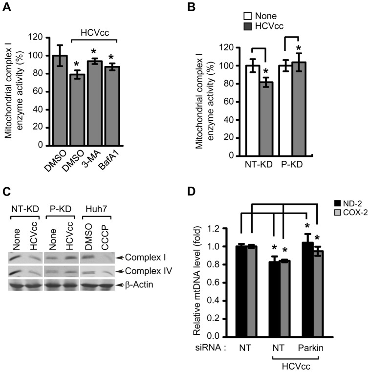
Figure 7. Mitochondrial functions altered by HCV infection are associated with Parkin-mediated mitophagy.
(A) Rescue effect of 3-MA and BafA1 on reduction of mitochondrial complex I enzyme activity caused by HCV infection. Huh7 cells infected with HCVcc were treated with 3-MA (10 mM) or BafA1 (100 nM) for 12 h before harvest. At 3 days post-infection, the activity of mitochondrial complex I enzyme was measured according to manufacturer's instructions (mean ± SD; n = 3; *p<0.01). (B) Rescue effect of Parkin knockdown on reduction of mitochondrial complex I enzyme activity caused by HCV infection. NT-KD and P-KD cells infected with HCVcc were harvested on day 3 post-infection and used for analysis of the activity of mitochondrial complex I enzyme (mean ± SD; n = 3; *p<0.05). (C) Western blot analysis of mitochondrial respiratory chain complex enzyme expression. NT-KD and P-KD cells were infected with HCVcc and at 3 days post-infection, the expression levels of complex I and IV enzyme were analyzed by immunoblotting with anti-Hu total OxPhos complex antibody. Huh7 cells treated with CCCP (10 µM) for 12 h were also analyzed as a control. β-actin was used as an internal loading control. P values were calculated by using an unpaired Student's t-test. (D) Effect of Parkin silencing on depletion of mitochondrial DNA caused by HCV infection. Huh7 cells transfected with NT or Parkin-specific siRNA pools were infected with HCVcc. At day 3 post-infection, mitochondrial DNA levels of ND-2 and COX-2 were analyzed by real-time qPCR. GAPDH was used to normalize changes in ND-2 and COX-2 gene expression (mean ± SD; n = 3; *p<0.01). (A, B, and D) P values were calculated by using an unpaired Student's t-test.

