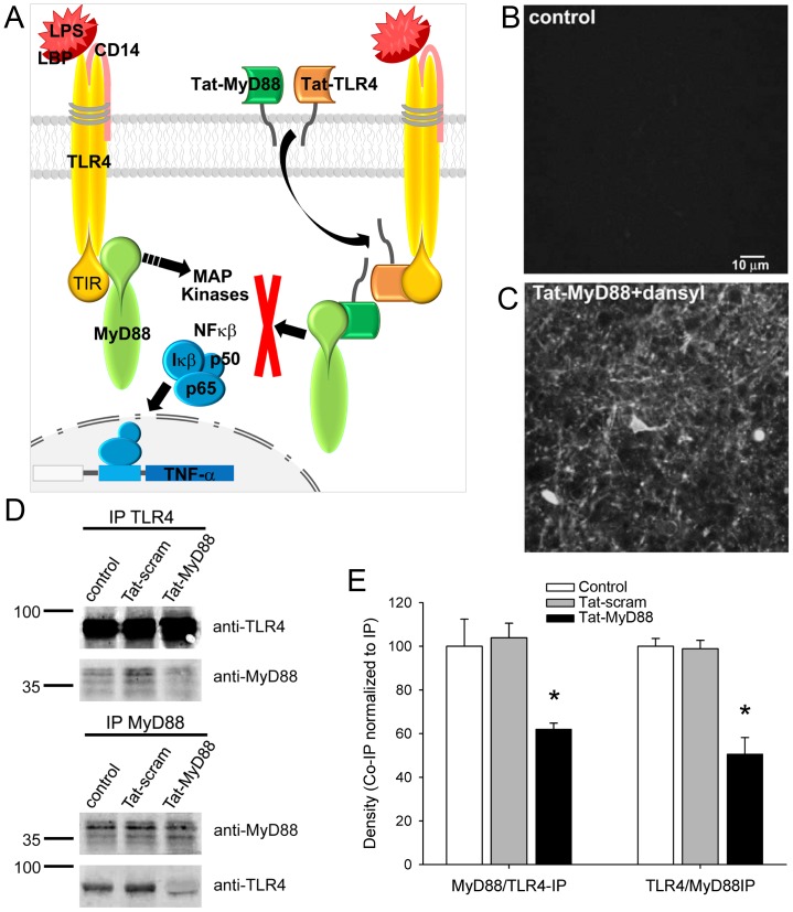
Figure 1. Schematic diagram showing the mechanism of action of the Tat-MyD88 and Tat-TLR4 peptides and their efficacy in preventing protein interactions in vivo.
A. The peptides are directed against regions of the TLR4 receptor and MyD88 TIR domain, thereby interfering with the interaction of these two proteins. LPS treatment has been shown to increase TLR4 and MyD88 binding leading to the activation of MAP kinases and NFκβ modulation of TNF-α. Thus the peptides may be effective in blocking downstream signalling to MAP kinases and TNF-α. B,C. 2-photon images of hippocampal tissue following intraperitoneal (i.p.) injection in the mouse reveals that dansylated Tat peptide can be observed in brain cells. D When i.p. injected, Tat-MyD88 but not Tat-scram reduced co-immunoprecipitation of TLR4 and MyD88 from brain tissue. E Densitometry quantification of co-immunoprecipitated protein normalized to immunoprecipitated protein.

