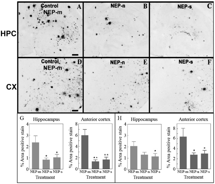
Figure 2. Study 1: Aβ immunostaining is observed in mice throughout both the ipsilateral hippocampus (A, B and C) and ipsilateral anterior cortex (D, E and F).
Aβ staining in the hippocampus of animals that received intracranial injections of rAAV- NEP-n (B) or NEP-s (C) is reduced compared to staining in those animals that received injections of control vector rAAV- NEP-m (A). Aβ staining in the anterior cortex of mice that received intracranial injections of rAAV- NEP-s (F) or NEP-n (E) is also reduced compared to staining in mice that received control vector rAAV- NEP-m (D). Scale bar = 120 µm. Quantification of percent area of positive total Aβ staining is shown in the hemisphere ipsilateral to injection sites (G) and in the hemisphere contralateral to injection sites (H). NEP-n (n = 18), NEP-m (n = 15), NEP-s (n = 17). The (*) indicates significance compared to NEP-m with p<0.05; the (**) indicate significance compared to NEP-m with p<.001.

