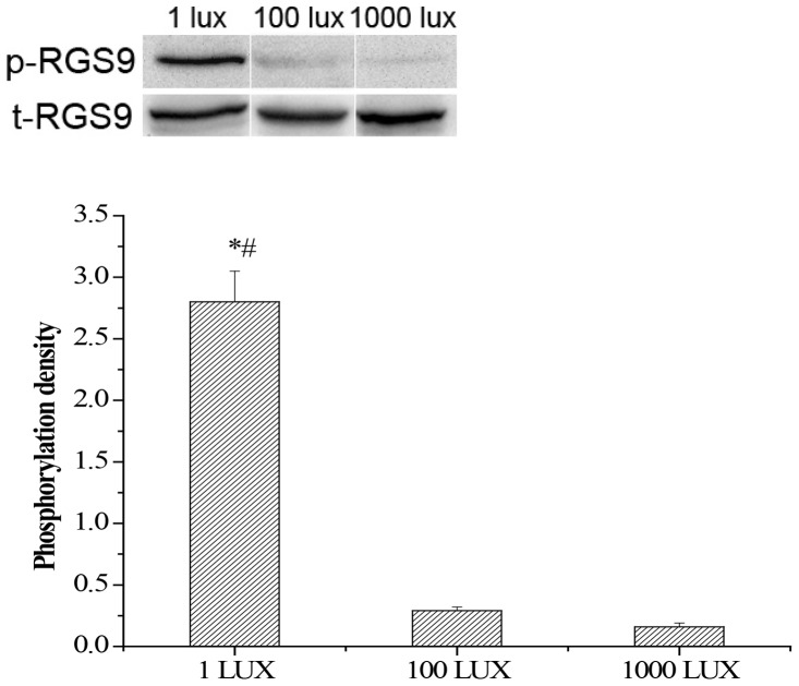
Figure 8. RGS9-1 is heavily phosphrylated on Ser475 under very dim light exposure.
Upper panel: Mouse retinas isolated under 1 lux, 100 lux and 1000 lux were analyzed by Western blotting probed with a anti-Ser475 phospho-RGS9-1 antibody (p-RGS9) and a anti-RGS9 antibody (t-RGS9). The total RGS9 (t-RGS9) is consistent under various light/dark conditions. Lower panel: Densitometric profiles of the densities of the Western blots of individual p-RGS9 bands. Densitometric scans of the autoradiographs were quantified and analyzed statistically. Error bars represent SEM, n = 5. Asterisk denotes statistically significant differences (p<0.05); # indicates significantly different than 1000 lux group at P<0.01 level.

