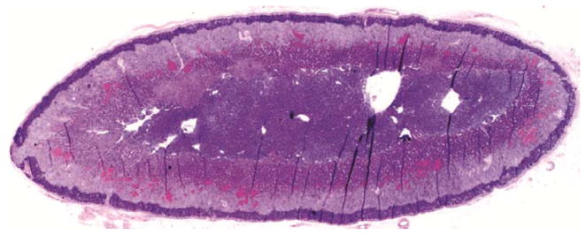
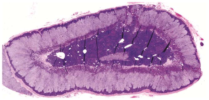
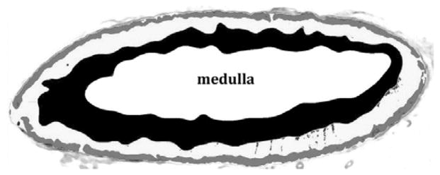
Figure 1.


Histological sections through rhesus adrenal glands. A. Hemotoxylin/eosin stained section of old female adrenal. B. Hemotoxylin/eosin stained section of young female adrenal. C. Processed image of adrenal section in B, showing delineation of the areas comprising the zona reticularis (black), zona glomerulosa (gray), intervening zona fasciculata (white) and central medulla (labeled).
