
Figure 2. IL-22−/− mice exhibit defects in the dermis during wound healing.
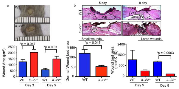
A) Images of WT and IL-22−/− backskin 3 days after wounding with 2mm punch biopsies. Quantification of the external area of WT and IL-22−/− small wounds at 3 and 5 days post-wounding. n>18 wounds for each genotype (6 wounds from 6 mice for each of 3 independent experiments). Ruler notches=1mm. B) Hematoxylin and eosin stained sections of WT and IL-22−/− backskins wounded with 8mm punch biopsies (large wounds). Epidermis (e) is indicated by dotted line and wound bed (WB) is outlined by solid line. Quantification of wound bed area (i.e. dermal area) of small wounds (2mm) 3 days post wounding and large wounds (8mm) 5 and 8 days post wounding in WT and IL-22−/− mice are shown. For small wounds, n = 18 (6 wounds from 6 mice for each of 3 independent experiments). For large wounds, n= 12 wounds from 6 mice. Bars = 400μm. All data are ± SEM. Asterisks indicate significance.