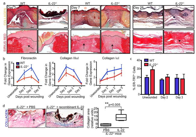
Figure 3. IL-22−/− mice exhibit defects in extracellular matrix production.
A) Skin sections of WT and IL-22−/− mice 3, 7 and 28 days post wounding with 2mm punch biopsies were stained with Movat Pentachrome (collagen, yellow; mucin, blue) and Sirius Red (Collagen Type III, red). Bars = 200 μm. Arrows indicate collagen, arrowhead indicates disorganized collagen. B) Real Time PCR analysis reveals decreased Fibronectin, Collagen IaI and Collagen IIIaI mRNA levels in 2mm IL-22−/− skin wounds. n= 4 mice for each genotype and timepoint. C) Quantification of % ER-TR7+ cells in the total dermal cell population derived from the dermis of WT and IL-22−/− mice wounded with 2mm punch biopsies. n=4 mice for each genotype. D) Skin sections of IL-22−/− skin 5 days post-wounding that were injected with PBS or recombinant IL-22 on day 3 and stained with trichrome histological stain (collagen, blue). Bars = 200 μm. Quantification of the area of collagen in the wound beds of IL-22−/− mice injected with PBS or 25ng recombinant IL-22. n= 6 mice. Asterisks indicate significance. All data are mean +/− SEM.

