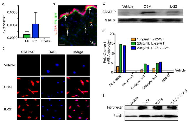
Figure 4. IL-22 can stimulate STAT3 activation and ECM production in dermal fibroblasts.
A) Real time PCR reveals that isolated dermal fibroblasts (FB) and keratinocytes (KC) express IL-22Rα mRNA. n=3 mice. Data are mean +/− SEM. B) Skin sections of were immunostained with antibodies against IL-22Rα and ER-TR7. Arrows indicate ER-TR7+ and IL-22Rα^Ecells. Bar = 100 μm. epi, epidermis. C) Representative images of primary mouse fibroblasts immunostained with antibodies against phospho-STAT3 (STAT3-P) after treated with IL-22 or oncostatin M (OSM) for 20 minutes. D) Western blot analysis confirms the increased activation of STAT3-P in primary dermal fibroblasts treated with IL-22 or Oncostatin M. E) Real time PCR analysis of mRNA in WT or IL-22−/− primary mouse fibroblasts reveals enhanced expression of ECM genes after IL-22 stimulation for 48 hours. F) Western blot analysis shows that IL-22 increases the expression of fibronectin protein in WT fibroblasts. β-actin is included as a loading control.

