
Figure 5. IL-22 induces myofibroblast differentiation.
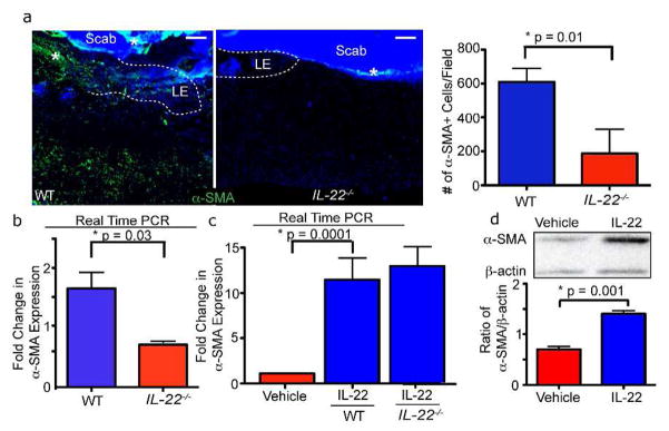
A) Skin sections of WT and IL-22−/− mice 3 days after wounding with 2mm punch biopsies were immunostained with antibodies against α-SMA. Quantification of α-SMA staining, n = 6 mice for each genotype. B) Real time PCR reveals reduced mRNA expression of α-SMA in 2mm IL-22−/− wounds 3 days post-wounding. n= 4 mice for each genotype and each timepoint. C) Real time PCR analysis of WT or IL-22−/− primary dermal fibroblasts cultured with vehicle or recombinant IL-22 for 48 hours confirms that α-SMA mRNA expression increases with IL-22 stimulation. n = 3 independent experiments. D) α-SMA protein expression increases in primary fibroblasts after stimulation with IL-22 for 48 hours as indicated by representative image of western analysis. Quantification of the ratio of α-SMA/β-actin expression is shown. n = 3 independent experiments. All data are mean +/− SEM.