
Figure 4.
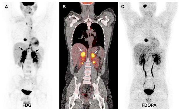
Multicentric SDHD-related PGL syndrome (1 HNPGL, 1 cardiac, 2 adrenal PHEO). A. 18F-FDG PET (MIP image) showing positive cardiac PGL and bilateral PHEO (arrows). Non-specific uptake in the mediastinum corresponds to brown fat. The HNPGL is not visible on this projection. B. Coronal fused 18F-FDG PET/CT image centered over the PHEO (2 positive tumours, arrows). C. 18F-FDOPA PET (MIP image) showing positive HNPGL and cardiac PGL, negative bilateral PHEO.