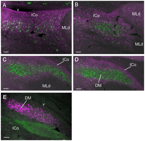
Figure 10.
A–D: Anterograde (red) and retrograde (green) labeling in ICo and DM following an injection of CTB 555 into POM and an injection of CTB 484 into RAm in the same case. Note how in A the retrogradely labeled cells are interspersed among the anterogradely labeled fibers and terminations, whereas in B–D they form the tightly clustered DM nucleus that does not admit the anterogradely labeled fibers, but is surrounded by them. E: DM (red) retrogradely labeled from an injection of CTB 555 in RAm, dorsal to and separate from a terminal field (green) in ICo resulting from an injection of CTB 484 into the medial arcopallium. Scale bar = 100 μm.

