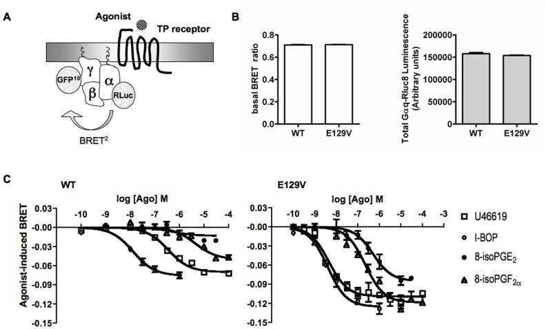
Figure 6. BRET2 measurement of Gαqβ1γ2 complex activation in HEK293 living cells expressing equal amounts of the WT of human TP receptor or its E129V mutant.
A. BRET2 was measured between the donor Rluc8 and the acceptor GFP10 introduced at the residue 97 of the Gαq subunit and the N-terminal domain of the Gγ2 subunit, respectively. Agonist-induced coupling of TP receptor and Gq protein distances Gαq-Rluc8 and GFP10-Gγ2 giving rise to a decrease in the BRET signal. B. Protein expression levels of the constructs used for BRET experiments were set to be constant and able to assure the same level of basal BRET ratio in the presence of WT and E129V mutant of the human TP receptors. Total Gαq-Rluc8 luminescence was evaluated in HEK293 cells co-expressing Gαq-Rluc8 together with GFP10-Gγ2 and Gβ1 in the presence of WT or E129V mutant of the human TP receptor measuring the light emission in aliquots of the transfected cells incubated with 5 µM coelenterazine for 8 min. In the same cells stimulated with PBS, basal BRET ratio was calculated as the ratio of the light emitted by GFP10 (510–540 nm) over the light emitted by Rluc8 (370–450 nm). C. BRET was measured in HEK293 cells co-expressing Gαq-Rluc8 together with GFP10-Gγ2 and Gβ1 in the presence of WT (left) or E129V (right) mutant of the human TP receptor and stimulated with increasing concentrations of the indicated full and partial agonists. Results are the differences in the BRET signal measured in the presence and the absence of agonists, and are expressed as the mean value±SE of at least two independent determinations.

