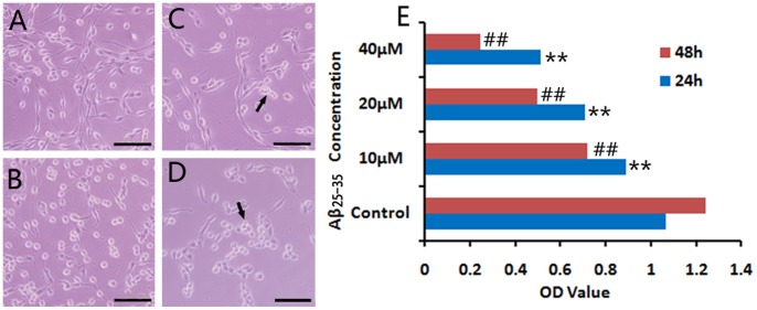
Figure 1. Survival of PC12 cells exposed to Aβ25–35.
PC12 cells were cultured for 24 and 48 h with 10, 20 and 40 µM Aβ25–35, respectively. After 24 h in vitro culture, (A) PC12 cells of the normal control group grew well with long neurites. (B) While incubation with 10 µM Aβ25–35, neurites of cells retracted gradually. (C) When added 20 µM Aβ25–35 in the medium, the damage to cells was obvious, and some cell debris appeared. (D) The 40 µM Aβ25–35 strongly insulted cells resulting in loss of more cells. After 48 h with Aβ25–35, more cells floated in the medium, and cell debris increased. Arrows indicated cell debris. Cell viability was assessed by MTT assay. Absorbance values were presented as mean ± S.E.M. **p < 0.01, compared with the normal control group at 24 h in culture; ## p < 0.01, compared with the normal control group at 48 h in culture. Data were processed by two-way ANOVA followed by Newman-Keuls test (n = 9). Scale bars: A-D, 100 µm.

