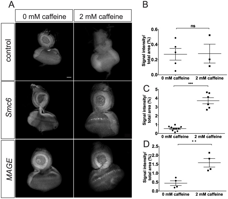
Figure 4. Caffeine exposure results in apoptosis in eye discs of MAGE and Smc6 mutants.
(A) Anti-cleaved-caspase-3 antibody staining of eye discs from third instar larvae of control (WT, FRT82B), MAGE (sstRZ/sstXL), and Smc6 (jnjX1/jnjR1) genotypes raised in either standard media (0 mM caffeine) or media supplemented with 2 mM caffeine for 12 hours before dissection. Images are single stacks of confocal images. More cleaved-caspase-3 foci in eye discs of sstRZ/sstXL and jnjX1/jnjR1 larvae were observed after caffeine exposure. A narrow band of apoptotic cells (white arrow heads) anterior to the presumptive morphogenetic furrow are most noticeable. Scale bar represents 50 µM. (B-D) Quantification and comparison of cleaved caspase-3 staining levels in WT (B), MAGE (C) or Smc6 (D) eye discs, comparing the no caffeine and 2 mM caffeine groups. Data represent mean area stained from multiple eye discs for each genotype per treatment. A maximum projection of all stacks of a confocal image was used to quantify the signal intensity of staining. This value was divided by the area of each eye disc to obtain a ratio representing the relative amount of immunostaining. Error bars represent SEM. A non-paired two-tailed t-test was used to determine statistical significance. **, P = 0.006, ***, P<0.0001.

