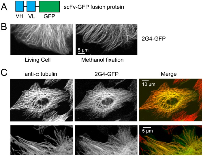
Figure 1. The scFv 2G4-GFP colocalizes with the majority of microtubules.
(A) Diagram outlining the recombinant antibody. The combined mw of the VH and VL regions approximately equals that of EGFP. (B) 2G4-GFP localizes to linear filaments in either living cells or in cells fixed in −20°C methanol. Images shown are from the edges of two different LLCPK cells (scale bar = 5 µm). (C) 2G4-GFP localizes to the majority of microtubules. LLCPKs were transfected with plasmid encoding 2G4-GFP and fixed 24 h later. Microtubules were stained with an antibody to a-tubulin. Images in the top row show a maximum intensity projection from a Z series (scale bar = 10 µm). The bottom row shows single optical section from the edge of a second LLCPK cell (scale bar = 5 µm).

