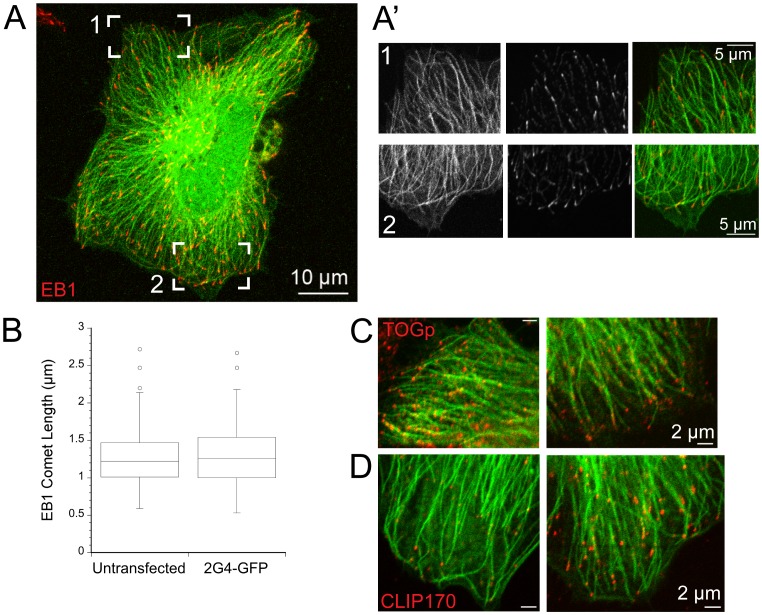
Figure 2. Expression of scFv 2G4-GFP does not disrupt protein binding to microtubule plus ends.
(A) 2G4-GFP binding extends to the distal ends of microtubules, as marked by EB1. A maximum intensity projection from optical sections through a Hela cell is shown (scale bar = 10 µm). (A′) Single optical sections from bracketed regions of the cell shown in (A) (scale bar = 5 µm). 2G4-GFP binds uniformly along microtubules and extends to the ends of these microtubules. (B) Box plot of EB1 comet lengths at microtubule plus ends. Expression of 2G4-GFP did not change the length of EB1 comets. (C) TOGp, another microtubule plus end binding protein, is localized to microtubules labeled by 2G4-GFP. (D) CLIP-170 was also localized to microtubule plus ends in cells expressing 2G4-GFP. Note that CLIP-170 was localized to only a subset of microtubule ends. We observed a similar pattern in Hela cells expressing GFP- a-tubulin (data not shown). Scale bars in C, D = 2 µm.

