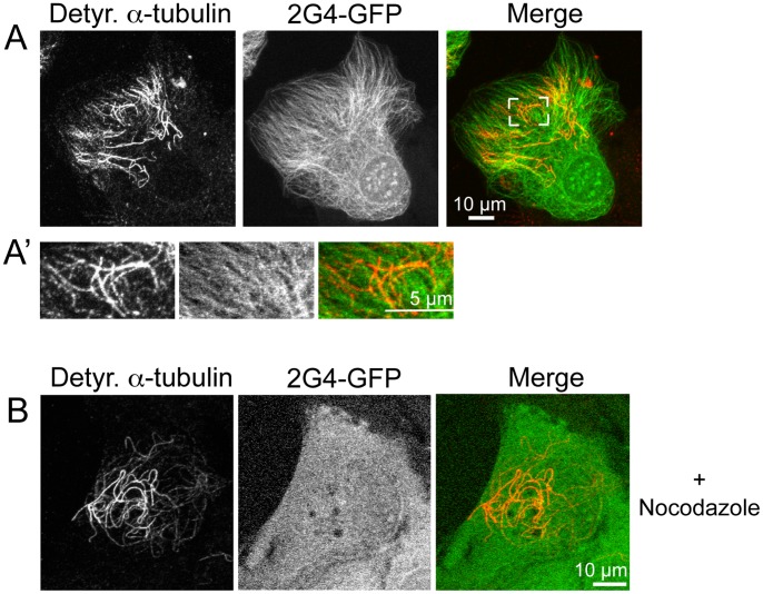
Figure 8. Recombinant scFv 2G4-GFP does not co-localize with de-tyrosinated α-tubulin.
(A) LLCPK cells were fixed 24 h after transfection and stained with antibodies specific for de-tyrosinated α-tubulin. The area bracketed in white is enlarged in (A′). 2G4-GFP does not show detectable binding to microtubules recognized by an antibody specific for detyrosinated α-tubulin. (B) LLCPK cells expressing 2G4-GFP were incubated in 33 µM nocodazole for 15 m prior to fixation and localization of detyrosinated α-tubulin. Depolymerization of the majority of microtubules shifted 2G4-GFP to a soluble protein present uniformly throughout the cell and it did not colocalize with microtubules composed of detyrosinated α-tubulin. Scale bars = 10 µm (whole cell images) and 5 µm (enlarged region). Images shown are maximum intensity projections from optical sections.

