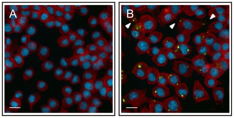
Figure 6. The morphology of macrophages changes following infection with R. equi.
J774A.1 monolayers were infected with ATTO 488 labelled virulent R. equi 103+ (green). Monolayers were fixed 24 h post-infection; the actin cytoskeleton and cell nuclei were stained with Texas Red Phalloidin (red) and Hoechst 33258 (blue), respectively. Panel A: non-infected monolayers, panel B: monolayers infected with R. equi 103+. Arrows indicate the change in cell shape following infection with R. equi. The length of the white bar is 40 µm.

