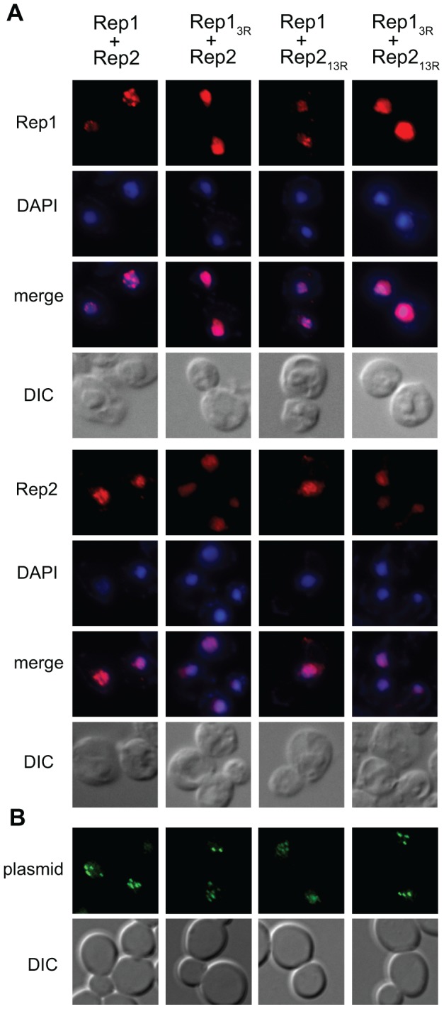
Figure 7. Localization of Rep13R and Rep213R.

(A) Spheroplasts were prepared from cir 0 yeast expressing wild-type or mutant Rep1 and Rep2 from an ADE2-tagged 2 µm plasmid and the Rep proteins were visualized by indirect immunofluorescence. Bulk chromatin was visualized by DAPI staining and cells by light microscopy (DIC). (B) Yeast were co-transformed with an ADE2-tagged 2 µm plasmid encoding the indicated Rep1 and Rep2 alleles and a plasmid containing the STB locus and 256 lacO repeats. Plasmid localization was visualized by fluorescence microscopy following induction of GFP-LacI repressor fusion protein expression.
