
FIGURE 1.
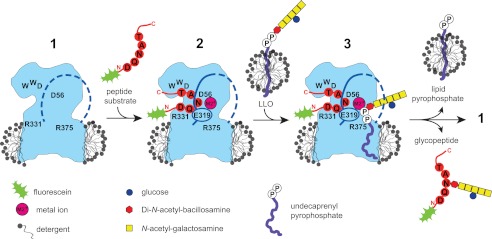
Schematic of a working model of the PglB reaction cycle. The contour of C. lari PglB in a detergent micelle is shown in blue, with positions of functionally relevant and conserved residues indicated in black. In the ground state (1), the external loop 5 (EL5) is assumed to be disordered (dashed blue line). Binding of a fluorescein-labeled substrate peptide and divalent metal cation (M2+, pink) causes EL5 (including residue Glu-319) to become ordered (continuous line) (2). Addition of extracted, LLO from a separate micelle triggers glycosylation, with state 3 likely reflecting a transient state. Following the glycosylation reaction, glycopeptide and undecaprenyl pyrophosphate are released.