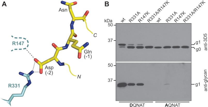
FIGURE 6.
Interactions between the −2 sequon position and PglB. A, ball and stick representation of acceptor peptide (yellow) and interacting PglB residues (cyan), as observed in the X-ray structure (PDB code 3RCE). Sequon and PglB residues are labeled in black and cyan, respectively. Yellow lines indicate N and C termini of the sequon. Proposed hydrogen bonds are shown as dashed lines. A potential interaction between Arg-147 and the −2 Asp is indicated. B, immunoblots of in vivo glycosylation reactions detecting acceptor protein 3D5 (top) or bacterial N-glycans (bottom). Glycosylation results in a mobility shift from the non-glycosylated (g0) to the glycosylated form of the acceptor protein (g1). PglB mutants are indicated above the lanes, the glycosylation sequons present in the acceptor proteins are indicated below the lanes.

