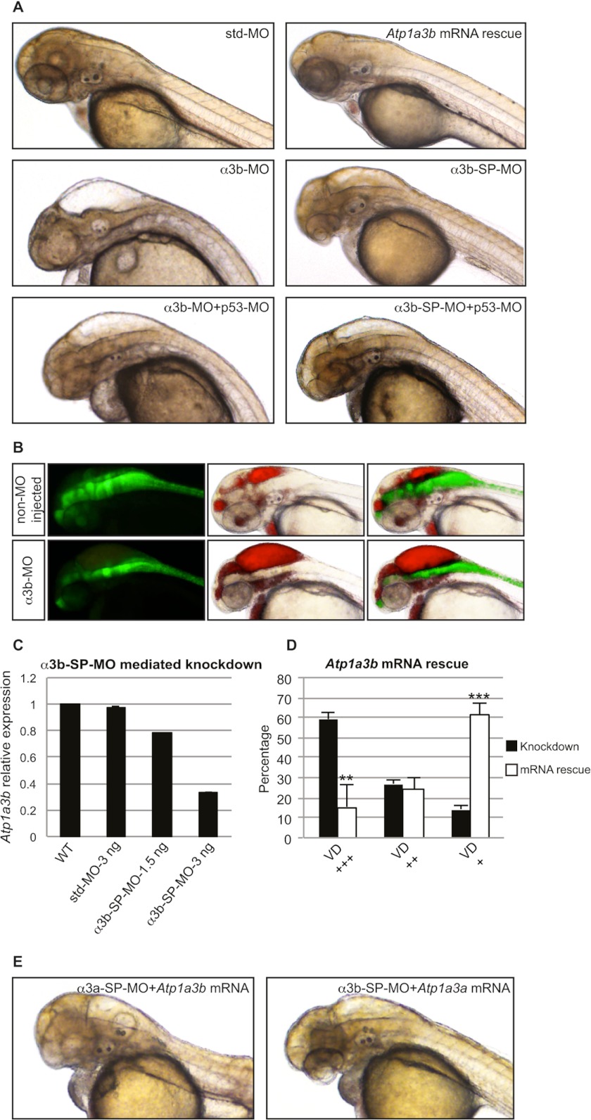
FIGURE 3.
Knockdown of Atp1a3b phenocopies Atp1a3a knockdown. A, brain ventricle dilation was observed in embryos upon Atp1a3b KD mediated by α3b-MO- or α3b-SP-MO-injected embryo as compared with the std-MO-injected control embryo. This phenotype was rescued by co-injection of Atp1a3b mRNA. p53-MO co-injections with any of the MOs did not rescue the brain ventricle dilation phenotype. B, brain ventricles of Tg(gfap:GFP) line, non-MO-injected and α3b-MO-injected, were injected with rhodamine-conjugated dextran. Brain ventricles and the astrocytes are highlighted by red and green fluorescence, respectively. C, α3b-SP-MO-mediated KD efficiency was tested by qRT-PCR in terms of changes in the relative expression level of Atp1a3b. Data are presented as mean ± S.E. of triplicate measurements. Concentrations of MOs are indicated. D, mean percentages ± S.D. of the α3b-SP-MO-injected embryos suffering from brain ventricle dilation (VD) of different severity, with (white columns, n = 148) and without (black columns, n = 88) Atp1a3a mRNA co-injection, are plotted. The degree of brain ventricle dilation is as follows: +, slight/no; ++, moderate; +++, severe. **, p < 0.01; ***, p < 0.001. E, Atp1a3a mRNA did not rescue α3b-SP-MO-injected embryos; similarly, Atp1a3b mRNA did not rescue α3a-SP-MO-injected embryos from brain ventricle dilation.

