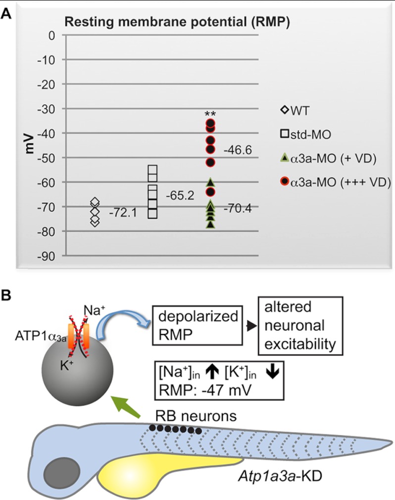
FIGURE 4.
RB neurons are more depolarized in Atp1a3a KD zebrafish displaying severe brain ventricle dilation. A, RMP values of RB neurons from WT (n = 5), std-MO-injected (n = 6), and α3a-MO-injected embryos (n = 10) are plotted. The α3a-MO-injected embryos are divided into embryos displaying severe (+++) (n = 5) or slight/no (+) (n = 5) brain ventricle dilation (VD). The number of cells (n) recorded per group stems from at least three different animals. RMP data are presented as mean ± S.D. **, p < 0.01 between RMPs of α3a-MO-injected embryos with severe ventricle dilation and control groups, WT, and std-MO-injected embryos. B, schematic representation summarizing the depolarization of the RMP in RB neurons in an Atp1a3a KD embryo. The scheme covers the time frame of 48–60 hpf. The dashed cross marks a malfunctioning α3aNa+/K+-ATPase.

