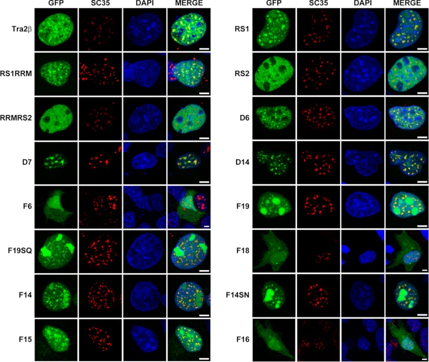
FIGURE 7.
Nuclear speckle distribution patterns of RS1, RS2 domain, truncations and mutants of GFP-RS1 in COS-1 cells. Confocal microscopy showing immunofluorescent signals of GFP (green), SC35 (red), DAPI (blue), and MERGE (overlay) in cells with expression of various GFP fusion proteins. Scale bars: 5 μm.

