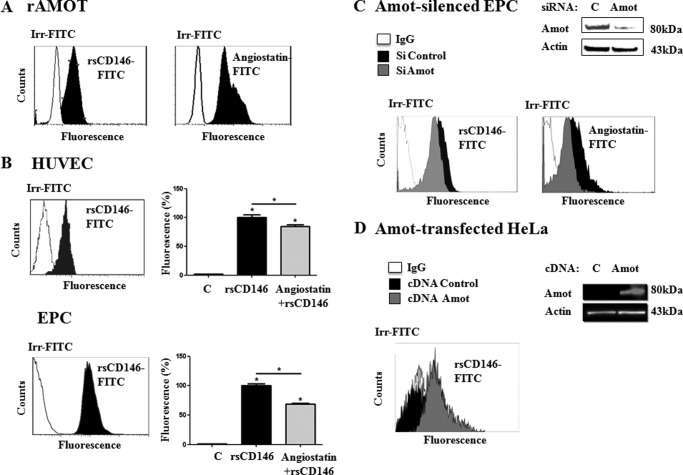
FIGURE 3.
Binding of angiomotin, angiostatin, and sCD146 on endothelial cells. A, binding of sCD146 on recombinant angiomotin. Recombinant angiomotin was coupled to Dynabeads through an anti-GST antibody (see “Experimental Procedures”). Interaction of the molecule with rsCD146-FITC (1.5 μm) or angiostatin (1.5 μm) coupled to anti-angiostatin antibody and secondary antibody was tested by flow cytometry. Isotypic control (Irr-FITC) was used. A representative experiment of three to five experiments is given. B, binding of sCD146 on HUVEC and EPC. Binding of rsCD146-FITC (1.5 μm) was observed on HUVEC or EPC by flow cytometry. Isotypic control (Irr-FITC) was used. The rsCD146-FITC binding was displaced by an equimolar concentration of angiostatin. A representative experiment is given for the flow cytometry graph and displacement by angiostatin is the average of three to five experiments. *, p < 0.05, experimental versus control. C, reduced sCD146 binding in angiomotin-silenced EPC. Binding of rsCD146-FITC (1.5 μm) was observed by flow cytometry in EPC silenced or not for angiomotin. Angiomotin silencing was visualized by Western blot. The binding of angiostatin (1.5 μm) was also tested. Isotypic control (Irr-FITC) was used. A representative experiment of three experiments is shown. D, enhanced sCD146 binding in angiomotin-transfected HeLa cells. Binding of rsCD146-FITC (1.5 μm) was observed by flow cytometry in HeLa cells transfected with angiomotin. Angiomotin was not expressed in wild-type HeLa but was expressed after angiomotin transfection, as visualized by Western blot. Isotypic control (Irr-FITC) was used. A representative experiment of three experiments is shown.

