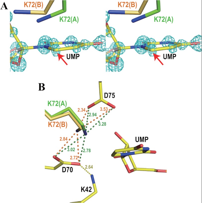
FIGURE 3.
Ligand binding site. A, stereo view of UMP and both conformations of Lys-72 superposed on the Fo − Fc omit electron density map of UMP contoured at 12 σ. Conformations A and B of Lys-72 are drawn in green and orange, respectively. A red arrow points to the C6 atom of UMP, which is displaced from the plane of the pyrimidine ring by 0.10 Å. B, the Lys-42-Asp-70-Lys-72-Asp-75′ network in the present atomic resolution structure. Numbers beside the dotted lines represent the distances between the two connected atoms in Angstroms. The color code is the same as in panel A.

