
FIGURE 4.
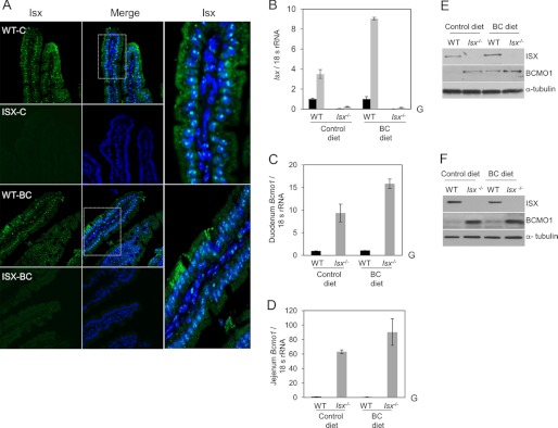
Consequences of ISX deficiency for BC metabolism. A, mouse duodenum from wild-type (WT) and Isx-deficient (Isx−/−) mice (n = 3 per genotype and diet) were immunostained with anti-ISX primary antibody, and ISX was visualized by using a secondary conjugated Alexa 488 antibody (green speckled fluorescence) and confocal microscopy. Nuclei were stained with DAPI. Representative images taken at ×40 magnification are shown. Speckled pattern of staining for ISX was observed in enterocyte nuclei. Approximately 20 sections per genotype and diet were scored for ISX localization. B–D, total RNA was isolated from intestinal tissue and expression of relevant genes was determined with gene-specific probe sets (ABI) and qRT-PCR. Values represent mean ± S.D. from 2 independent experiments carried out in triplicate. G, genotype effect in two way analysis of variance analysis (p < 0.05). B, solid black bars indicate the duodenal and gray bars indicate the jejunal mRNA expression levels. E and F, protein extracts from mice (n = 5 per genotype and diet) were obtained from intestinal tissue (duodenum and jejunum, respectively), pooled together, and 25 μg of protein per lane was subjected to immunoblot analysis. Immunoblots were probed with the relevant antibodies as indicated. α-Tubulin was used as the protein loading control. Representative immunoblots from three separate analysis are shown. WT, wild-type mice; Isx−/−, Isx deficient mice; BC diet, β-carotene diet.