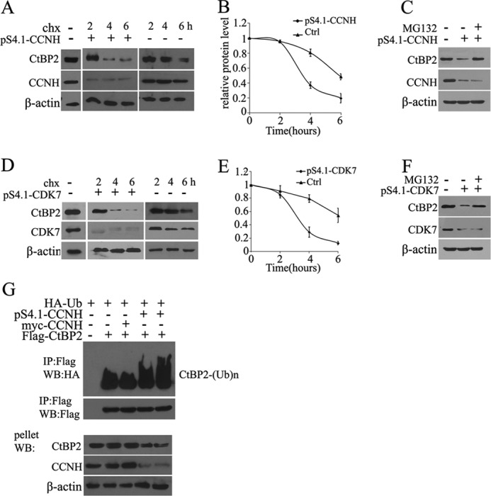
FIGURE 3.

Knockdown of CCNH or CDK7 causes CtBP2 degradation. A and B, HEK293T cells were transfected with the empty vector or pS4.1-CCNH (A), and 40 h later incubated with cycloheximide (chx). Cells were lysed at the indicated times (in hours) after cycloheximide addition and Western blotted with CtBP2 antibody. The relative amount of protein for CtBP2 is plotted below. C and D, 293T cells were transfected with the empty vector or pS4.1-CDK7 and 40 h later incubated with cycloheximide. Cells were lysed at the indicated times (in hours) after cycloheximide addition and Western blotted with CtBP2 antibody. The relative amount of protein for CTBP2 is plotted below. E and F, cells were treated and analyzed as in A or C, except that 30 min prior to the addition of cycloheximide, MG-132 was added to inhibit the proteasome. G, HEK293T cells were transfected with FLAG-tagged CtBP2, myc-tagged CCNH or pS4.1-CCNH, together with a HA-tagged ubiquitin expression construct, as indicated. FLAG immunoprecipitates or the pellet fraction was analyzed by FLAG and HA Western blotting (WB) as indicated.
