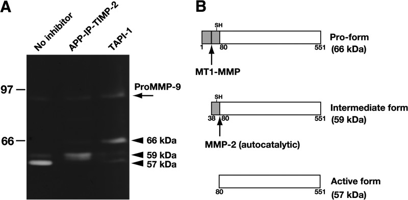
FIGURE 4.
Effect of APP-IP-TIMP-2 on processing of pro-MMP-2 in Con A-stimulated HT1080 cells. A, HT1080 cells were incubated for 24 h in serum-free medium containing Con A (100 μg/ml) with APP-IP-TIMP-2 (70 nm) or TAPI-1 (10 μm), or without inhibitor. The CMs were prepared from the incubated cells and subjected to gelatin zymography as described under “Experimental Procedures.” Arrowheads indicate the gelatinolytic bands of pro-MMP-2 at 66 kDa (upper), the intermediate form at 59 kDa (center), and the active form of MMP-2 at 57 kDa (lower). An arrow at 90 kDa indicates a gelatinolytic band of pro-MMP-9. Ordinate, molecular size in kDa. B, pro-MMP-2 (pro-form), the intermediate form, and the active form of MMP-2 are schematically represented. The numbers shown at the bottom of each scheme represent the amino acid residue numbers of pro-MMP-2. The MT1-MMP arrow and the MMP-2 arrow represent the sites of cleavage by MT1-MMP and MMP-2, respectively. SH represents the Cys residue interacting with catalytic zinc ion in pro-MMP-2.

