FIGURE 5.
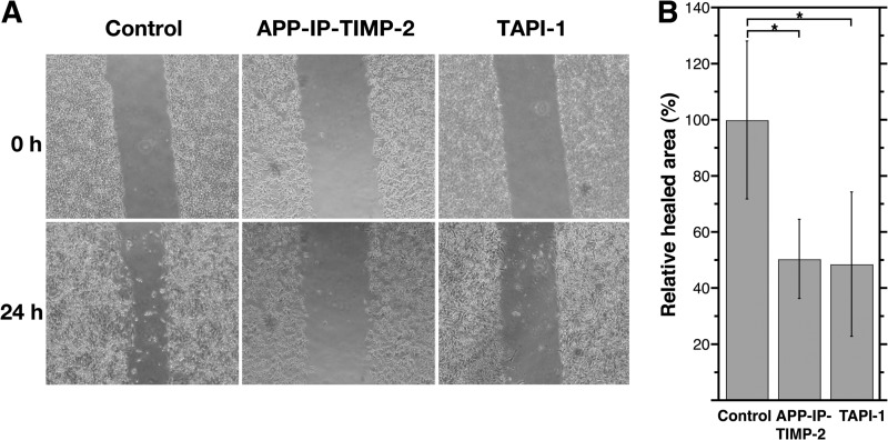
Effects of APP-IP-TIMP-2 on migration of Con A-stimulated HT1080 cells. HT1080 cells were cultured in 35-mm dishes until they reached confluence. Cells were treated for 30 min with serum-free DME/F-12 medium containing mitomycin C (25 μg/ml) and Con A (100 μg/ml). Scratch wounds were then made in the confluent monolayer. The wounded cell cultures were then incubated at 37 °C for 24 h in serum-free medium containing 70 nm APP-IP-TIMP-2 or 10 μm TAPI-1, or without inhibitor (Control). Before (0 h) or after (24 h) incubation, photographs were taken (A). The extent of cell migration in the presence or absence of inhibitors was quantified by measuring the areas of cells invading the scratch wounds using NIH Image J (B). In B, the area of invaded cells (healed area) in the absence of inhibitor was taken as 100%. The extent of cell migration is shown as the relative healed area on the ordinate. Each bar represents the mean ± S.D. for six assays. Statistical significance was determined by an unpaired test. *, p < 0.01.

