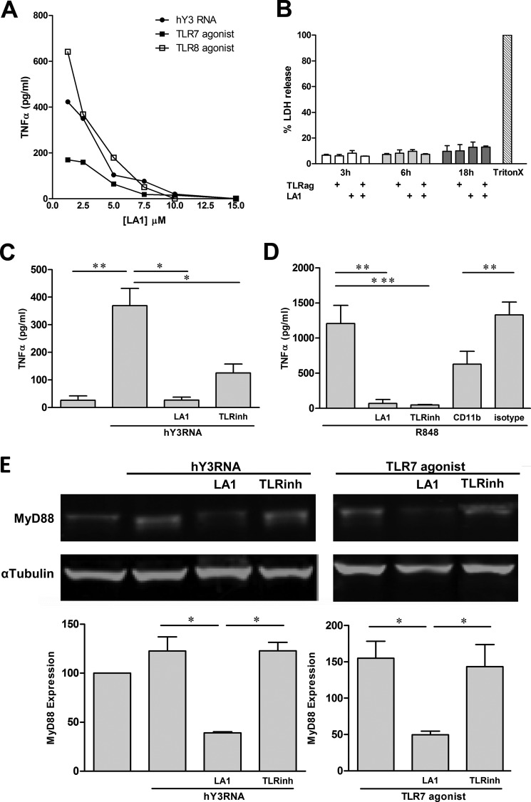
FIGURE 1.
LA1, a CR3 agonist, attenuates TNFα release by THP-1 cells stimulated with hY3, TLR7, and TLR8 agonists via the degradation of MyD88. A, TNFα levels measured in supernatants generated from THP-1 after exposure to either hY3 RNA transfection, a TLR7 agonist, and a TLR8 agonist (18 h, 37 °C) in the presence of LA1 (pretreatment of 30 min, concentration varied). B, lactate dehydrogenase (LDH) release relative to a positive control (a condition employing lysis using Triton X-100 (TritonX)) in supernatants from THP-1 cells stimulated with a TLR7/8 agonist for 3, 6, and 18 h with or without LA1. TNFα levels from THP-1 cells stimulated with hY3 (C) or R848 (D) in the presence or absence of LA1; an oligonucleotided-based TLR7/8/9 inhibitor (TLRinh); an anti-CD11b antibody; or isotype control. Bars represent mean pg/ml TNFα ± S.E. (A, n = 6; B, n = 4). E, immunoblot analysis of MyD88 in lysates generated from THP-1 cells stimulated with hY3 (left) or a TLR7 agonist (right) in the presence or absence of LA1 or a TLR7/8/9 inhibitor. α-Tubulin serves as a loading control. Bars represent the ratio of MyD88 to α-tubulin for each condition (mean value ±S.E. (n = 3). *, p < 0.05, **, p ≤ 0.005; ***, p ≤ 0.0005.

