FIGURE 1.
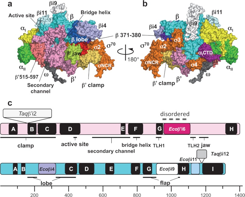
Three-dimensional crystal structure of the E. coli RNAP σ70 holoenzyme. a and b, surface representation of the E. coli RNAP holoenzyme. Panel a shows a view from the RNAP secondary channel leading to the active site, and panel b shows the σ-binding site. Each subunit of RNAP is denoted by a unique color: yellow, αI; green, αII; cyan, β; pink, β′; gray, ω; and orange, σ70. Several domains described under “Results and Discussion” are also denoted by a unique color and are indicated. c, linear maps of the β′ (upper) and β (lower) subunits. Conserved regions of the β′ subunit (A–H) and the β subunit (A–I) are shown as black boxes with the structural domains of RNAP. Specific insertions of the E. coli and Thermus RNAPs are shown by the same colors as in panels a and b and in Fig. 2.

